
Rewritten on: July 28, 2025
This article explains in detail how ultrasound (echo) screening during prenatal checkups is used to screen for Down syndrome, the significance of NT measurement, and its accuracy and limitations. It also covers the process of follow-up testing, such as NIPT, after a high-risk result.
What Is an Ultrasound (Echo) Scan?
 An ultrasound (echo) scan is a basic examination that everyone undergoes once pregnancy is confirmed. It is performed to check the growth and development of the fetus, gestational age, and the presence of any congenital abnormalities. It is a technology that uses sound waves to produce real-time images of the inside of the body, and because it does not use radiation (such as X-rays), it is a notable feature that it can be safely repeated for both mother and fetus.[ref:1]
Ultrasound scans play an extremely important role during pregnancy and are performed regularly to assess fetal development and the condition of the mother. Generally, prenatal checkups include multiple ultrasound scans across the early, middle, and late stages of pregnancy, with different points to check at each stage. Because it helps with early detection of abnormalities and risk management, it is an essential examination for pregnant women, and it is also used as a screening test for signs of genetic disorders and chromosomal abnormalities.
There are two main types of ultrasound scans: "transvaginal ultrasound" and "transabdominal ultrasound." In early pregnancy (up to about 12 weeks), the transvaginal method is mainly used, and the first things checked are whether a gestational sac can be seen in the uterus and whether a heartbeat can be confirmed. From the middle of pregnancy onward, the method switches to transabdominal, making it possible to observe the shape and development of each fetal organ in more detail.[ref:2] In recent years, 3D and 4D ultrasound technology has also advanced, making it possible to observe the three-dimensional shape of the fetus in real time. However, for Down syndrome screening, measurement in the conventional 2D mid-sagittal plane is still considered the most important method.[ref:8]
How Ultrasound Screening Assesses Down Syndrome Risk
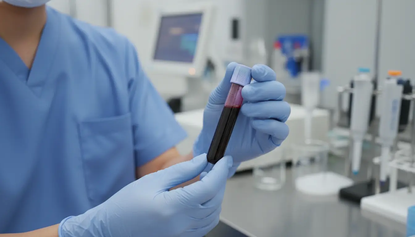 Ultrasound screening draws attention as a method for screening for Down syndrome (Trisomy 21) largely because of the measurement of NT (Nuchal Translucency: the fluid-filled space at the back of the fetus's neck), taken especially in early pregnancy, which is closely related to assessing Down syndrome risk. NT refers to the area of fluid buildup observed at the back of the fetus's neck. It is present to some degree in all fetuses, but if this thickness is larger than normal, it suggests an increased risk of chromosomal abnormalities, including Down syndrome.[ref:3]
NT measurement is generally performed between 11 weeks 0 days and 13 weeks 6 days of pregnancy, and a crown-rump length (CRL) of 45mm to 84mm is a condition for the measurement. The NT reference value varies depending on gestational age, but generally, a value of 3.0mm or more is considered "elevated," and further testing for chromosomal abnormalities may be recommended. However, a thick NT does not necessarily mean there is a chromosomal abnormality. Elevated NT can also occur in normal fetuses, and it is important to understand that it is merely one risk indicator among others.
In addition to NT measurement in early pregnancy, the following findings may also be used in assessing Down syndrome risk.
- Underdeveloped nasal bone (hypoplasia or absence) — If the nasal bone cannot be confirmed on an early ultrasound, the risk of Down syndrome is said to increase. One large-scale study reported that nasal bone hypoplasia was observed in about 60-70% of fetuses with Down syndrome.[ref:9]
- Structural heart abnormalities — Ventricular septal defects and atrioventricular septal defects are known to be heart conditions associated with Down syndrome
- Duodenal atresia (double bubble sign) — This may be observed on ultrasound from mid-pregnancy onward and has been noted to be associated with Down syndrome
- Shortened limbs — If the femur or humerus is shorter than expected for the gestational age, it is evaluated as one risk factor
- Echogenic bowel — A finding in which the intestines appear as bright as bone on ultrasound, considered a marker for chromosomal abnormalities including Down syndrome
These findings are called "soft markers." On their own, they do not provide grounds for diagnosis, but when several markers are observed at the same time, the risk is considered to be higher. In recent years, algorithms that combine the presence of these soft markers with maternal age, NT value, and serum markers to statistically calculate Down syndrome risk have become widely used.
Accuracy and Limitations of Ultrasound Screening for Detecting Down Syndrome
 Unlike Non-Invasive Prenatal Testing (NIPT), ultrasound screening has no age restrictions and can be undergone by all pregnant women. In particular, when used specifically to assess Down syndrome risk, it is common to conduct it as a combined screening test consisting of two parts — a blood test and an ultrasound scan — during early pregnancy.
One medical facility in Australia offers a blood test measuring two hormones (beta human chorionic gonadotropin and placental growth hormone) and one protein (pregnancy-associated plasma protein A: PAPP-A) together with an ultrasound scan between 12 and 13 weeks of pregnancy, and reports that this achieves a Down syndrome detection accuracy of approximately 85-90%. This detection rate is a figure for the "combined test" that integrates ultrasound and blood testing, and the accuracy of ultrasound screening alone is often lower than this.[ref:5]
On the other hand, according to data compiled by a major American genetic testing company, of 18 pregnant women diagnosed as high-risk by ultrasound screening, only 1 actually had a baby with Down syndrome, according to one report. In other words, the remaining 17 were "false positives," which is one of the major challenges of ultrasound screening.[ref:3]
The false-positive rate in combined screening tests is generally considered to be around 5%, and the positive predictive value (PPV) varies greatly depending on maternal age and other risk factors. In particular, for younger pregnant women, because the prevalence of Down syndrome is lower, there is a higher probability that a positive test result does not actually indicate Down syndrome.
The accuracy of ultrasound screening varies considerably, and there is not much data available on accuracy at domestic medical institutions either, but one thing that can be said is that because ultrasound screening is a screening test, additional testing is needed to accurately confirm the presence or absence of a condition. Factors that affect the accuracy of ultrasound screening include the following.
- Timing of the test — NT measurement must be performed between 11 and 13 weeks of pregnancy, and accurate measurement is not possible if this window is missed
- Skill and experience of the sonographer — Because NT measurement requires advanced technique, results can be affected by the sonographer's level of expertise. The UK's Fetal Medicine Foundation certifies sonographers' skills through an NT measurement quality assurance program, which is widely recognized as an international standard.[ref:10]
- Fetal position — Depending on the fetus's orientation and posture during the scan, accurate measurement can be difficult
- Maternal body type — The thickness of the abdominal wall, among other factors, can affect image clarity
- Performance of the equipment used — The resolution and performance of the ultrasound device also affects the accuracy of observable findings
What to Do If Ultrasound Screening Shows a High Risk
Even if ultrasound screening results in a "high risk" determination, this does not immediately confirm Down syndrome. As mentioned above, since this test has a high rate of false positives, it is important to calmly consider the next step.[ref:6]
Before NIPT was introduced in Japan in 2013, invasive tests such as amniocentesis and chorionic villus sampling were necessary, but with the development of NIPT, it became possible to perform a non-invasive test that is safe for both mother and fetus. Amniocentesis and chorionic villus sampling carry about a 1% risk of miscarriage and a risk of permanent injury to the fetus, but NIPT analyzes fetal DNA circulating in the mother's blood, making it 100% safe for both mother and fetus, and it can detect Down syndrome with an accuracy of over 95%.[ref:7]
Like ultrasound screening, NIPT is a screening test that requires a separate invasive definitive diagnosis to accurately confirm the presence or absence of the condition in question, but it is widely used as a follow-up test when an abnormality is found on ultrasound screening. The general process from a high-risk ultrasound result to a definitive diagnosis is as follows.
| Stage | Test Name | Characteristics |
|---|---|---|
| Stage 1 | Ultrasound Screening (Echo) | Non-invasive screening test. Relatively high false-positive rate |
| Stage 2 | NIPT (Non-Invasive Prenatal Testing) | Analyzes fetal DNA from maternal blood. High accuracy and non-invasive |
| Stage 3 | Amniocentesis / Chorionic Villus Sampling | Enables a definitive diagnosis. Carries a slight risk of miscarriage |
In this process, NIPT plays an important role as a bridge between ultrasound screening and a definitive (invasive) diagnosis. If NIPT results are negative, invasive testing can often be avoided, preventing the mother from taking on unnecessary miscarriage risk. In fact, multiple studies have shown that the introduction of NIPT has significantly reduced the number of invasive tests performed.[ref:8]
The Difference Between NIPT and Ultrasound Screening
Both NIPT and ultrasound screening are non-invasive screening tests, but there are major differences in how they work and in their accuracy. Ultrasound screening uses sound waves to observe the fetus's morphological features, whereas NIPT analyzes cfDNA (cell-free DNA) present in maternal blood to detect numerical chromosomal abnormalities.
NIPT's sensitivity for Down syndrome (Trisomy 21) is over 99%, its specificity is also over 99%, and its false-positive rate has been reported to be less than 0.1% — all extremely high figures. In comparison, the sensitivity of ultrasound screening alone is said to be around 60-70%, and even the combined test only reaches 85-90%. Because of this difference in accuracy, in recent years more and more medical institutions have tended to recommend NIPT as a follow-up test when a concerning finding is discovered on ultrasound screening.[ref:5]
However, it is important to understand that NIPT and ultrasound screening are not competing tests, but complementary tests that each play a different role. NIPT specializes in detecting numerical chromosomal abnormalities (trisomy 21, 18, 13, etc.), but morphological abnormalities in the fetus (such as structural heart defects or organ development) can only be confirmed by ultrasound screening. Therefore, even if NIPT results are negative, regular ultrasound evaluation of the fetus's morphology continues to be important.
seeDNA offers a campaign in which NIPT can be performed for free at the same time as prenatal DNA testing that confirms the biological parent-child relationship between the unborn baby and the father. There are no additional fees, no need for additional blood draws, and no extension of the testing period whatsoever. Please leave your genetic testing needs during pregnancy to seeDNA.
Sex can also be determined
Frequently Asked Questions
Q1. Can ultrasound screening alone provide a definitive diagnosis of Down syndrome?
A. No, ultrasound screening is only a screening test and cannot provide a definitive diagnosis of Down syndrome. If ultrasound screening results in a "high risk" determination, this must be confirmed through additional testing such as NIPT or amniocentesis. It is not medically recommended to make a final judgment based solely on ultrasound results.[ref:1]
Q2. If the NT (nuchal fold thickness) is thick, does that always mean Down syndrome?
A. No, a thick NT does not necessarily mean Down syndrome. Elevated NT is also seen in a certain proportion of normal fetuses. NT is merely one risk indicator, and even when the NT value is higher than the reference range, most fetuses are found to be healthy in many cases.[ref:3]
Q3. If I'm told my ultrasound result is high-risk, what test should I get next?
A. If ultrasound screening results in a high-risk determination, NIPT (Non-Invasive Prenatal Testing) is often recommended as the next step. NIPT is a non-invasive test that analyzes fetal DNA from maternal blood, and boasts extremely high accuracy, with a Down syndrome detection sensitivity of over 99%. If the NIPT result is positive, it is common to proceed to a definitive diagnostic test such as amniocentesis.[ref:5]
Q4. What is the Down syndrome detection rate for ultrasound screening?
A. The Down syndrome detection rate for ultrasound screening alone is said to be around 60-70%. When combined with a blood test as a combined screening test, a detection rate of 85-90% has been reported. However, because the false-positive rate is relatively high at around 5%, it should be noted that even when a high-risk determination is given, the fetus often does not actually have Down syndrome.[ref:5]
Q5. Which is more accurate, NIPT or ultrasound screening?
A. For detecting Down syndrome (Trisomy 21), NIPT is far more accurate. NIPT has a sensitivity of over 99% and a false-positive rate of less than 0.1%, whereas combined screening tests that include ultrasound have a sensitivity of 85-90% and a false-positive rate of around 5%. For this reason, if a concerning finding is discovered on ultrasound screening, it is recommended to undergo NIPT as a follow-up test.[ref:8]
Q6. From when can I take seeDNA's NIPT?
A. seeDNA's NIPT can be taken from 10 weeks of pregnancy onward. We are also running a campaign that allows you to take NIPT for free at the same time as prenatal DNA testing, with no additional blood draw or fees required whatsoever. For details, please contact seeDNA's toll-free number (0120-919-097).
Peace of Mind Support from the seeDNA Genetic Medicine Research Institute
The seeDNA Genetic Medicine Research Institute is a trusted specialist institution for DNA testing and genetic testing that holds the international quality standard ISO9001 and the Privacy Mark for personal information protection.
If you are troubled by questions of blood relations between family members or parents and children, or a partner's infidelity, our DNA testing experts are here to support you so you can feel at ease — please feel free to contact us.
[Free Consultation with Specialist Staff]

If you have any questions,
please feel free to contact our toll-free number.
\ Open every day, including weekends /
Business hours: Monday-Sunday 9:00 AM-6:00 PM
(excluding public holidays)
 Author
Dr. Kihan Tomikane, M.D., Ph.D.
Graduated from the University of Tsukuba Graduate School, Master's/Doctoral Program in Biosystem Studies and Molecular Medicine
In 2017, developed the first prenatal DNA testing method in Japan(Patent 7331325) using trace DNA analysis technology(Patent 7121440)
[References]
(2) What Is Fetal Ultrasound Screening | Prenatal Testing Certification System Operations Committee
(3) Demography, August 2015
(4) Understanding Noninvasive Prenatal Testing (NIPT), April 2022
(5) Mol Carcinog, October 2001
(6) Sydney Ultrasound for Women, September 2019
(7) Nikkei Medical, December 2008
(8) Ann Thorac Surg, August 2014
(9) seeDNA Genetic Testing & DNA Testing