
Rewritten on: September 1, 2024
DNA is the genetic material that serves as the blueprint for living organisms and has a double helix structure, but what scientists actually see is a blurry image such as an X-ray diffraction pattern. This article explains the structure, form, and historical discovery of DNA in detail.
- ・What Is DNA?
- └ The Basic Structure of DNA and the Four Bases
- ・The Shape of DNA
- ・The True Shape of DNA — Photo 51 and X-Ray Diffraction
- └ The History of the Discovery of the Double Helix Structure
- └ Why the True Shape of DNA Is Hard to See
- └ The Biological Significance of DNA's Shape
- └ Applications in DNA Testing
What Is DNA?
 DNA stands for "Deoxyribonucleic Acid," and it is the genetic material that serves as the blueprint not only for humans but for many living organisms, including plants, insects, and animals [ref:1]. DNA exists within the nucleus of cells and encodes the information needed for protein synthesis. In humans, a DNA sequence of about 3 billion base pairs makes up the entire genome, and all genetic traits are recorded within this vast amount of information [ref:2].
By analyzing DNA, it is possible to assess the risk of developing certain diseases and to provide appropriate treatment and prevention. With recent advances in genomic medicine, DNA analysis technology has become indispensable in fields such as early cancer detection, diagnosis of hereditary diseases, and even personalized medicine (precision medicine).
In addition, DNA typing—which extracts DNA from cells that make up the body and analyzes its sequence to determine parent-child relationships and sibling relationships—is also widely practiced. DNA typing is used not only in the field of forensic science but also in confirming family relationships and in immigration procedures, making it one of the technologies indispensable to modern society.
The Basic Structure of DNA and the Four Bases
DNA is a macromolecular compound made up of basic units called "nucleotides" linked together like a chain. Each nucleotide consists of a sugar (deoxyribose), a phosphate group, and one of four types of bases (adenine: A, thymine: T, guanine: G, cytosine: C) [ref:3]. The order in which these four bases are arranged (the sequence) is genetic information itself, and is sometimes called "the code of life."
The bases follow a specific pairing rule (complementarity): adenine always bonds with thymine, and guanine always bonds with cytosine. This principle of complementary base pairing forms the foundation of the mechanism by which DNA is accurately replicated. Each time a cell divides, DNA is faithfully copied, and genetic information is passed on to the next generation of cells.
- Adenine (A) pairs with thymine (T) via hydrogen bonds
- Guanine (G) pairs with cytosine (C) via hydrogen bonds
- The sequence pattern of the four bases determines genetic information
- The human genome consists of approximately 3 billion base pairs
- Slight differences in DNA sequence create genetic diversity among individuals
The Shape of DNA
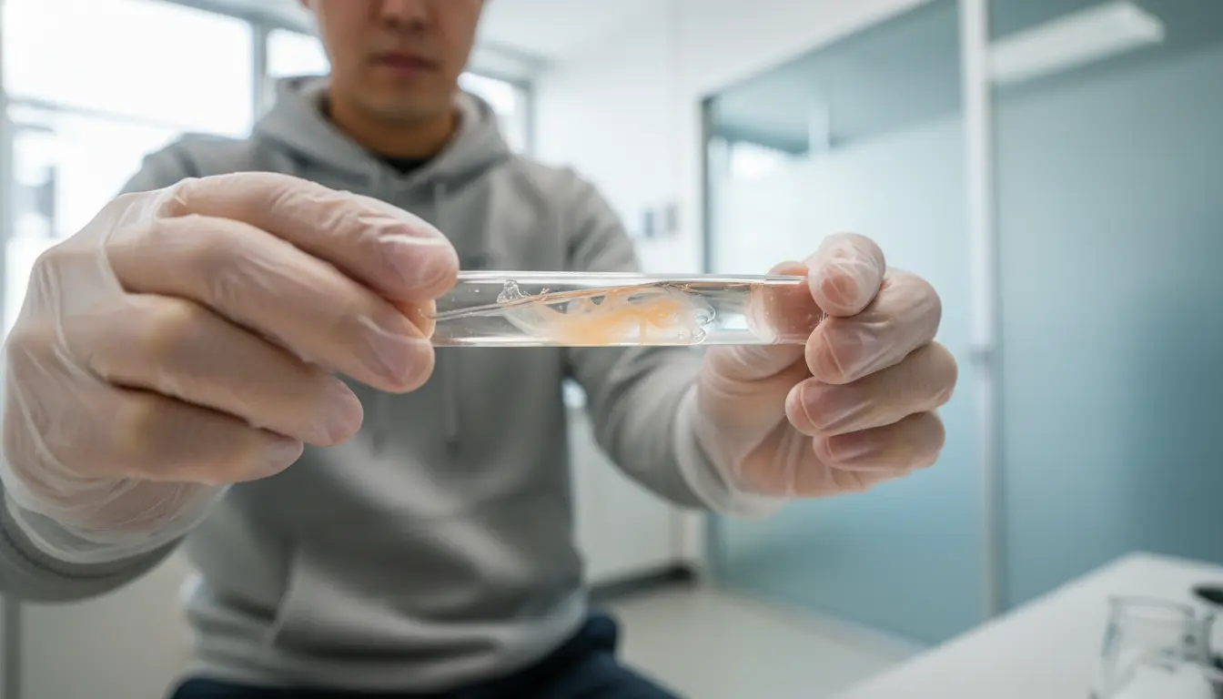 More than 20 years have already passed since the completion of the Human Genome Project, which read the entire human DNA sequence, and now even elementary school students picture a double helix when they think of the shape of DNA [ref:4]. That beautifully twisted image of two intertwined strands, which appears repeatedly in textbooks and the media, is one of the most iconic symbols of modern scientific literacy.
However, no one—including researchers who worked with DNA 20 years ago—has ever actually "seen" DNA looking like that clean computer-graphic image. The image of the double helix structure is nothing more than a "model" derived mathematically from experimental data; it is not the visible entity itself.
What scientists actually see is a blurry image like the one shown below. This fact is a crucial point for understanding the difference between "observation" and "modeling" in science.
The True Shape of DNA — Photo 51 and X-Ray Diffraction
 The blurry image shown above is an "X-ray diffraction image" obtained by irradiating crystallized DNA with X-rays. This technique, in which X-rays similar to those used to view human bones are directed at a crystal and the resulting diffraction pattern is recorded on photographic film, is called "X-ray crystallography" [ref:5].
The most famous example is the X-ray diffraction image known by the nickname "Photo 51." Taken in 1952 by Rosalind Franklin and Raymond Gosling, this image became decisive evidence that DNA has a helical structure [ref:5]. The characteristic X-shaped pattern captured in Photo 51 suggested that DNA had a helical structure, and by analyzing it using a complex mathematical method (Fourier transform), the now-famous double helix model was derived.
The History of the Discovery of the Double Helix Structure
In 1953, James Watson and Francis Crick proposed the double helix model of DNA based on experimental data including Photo 51. This discovery is regarded as one of the greatest achievements in the history of science and later led to the awarding of the Nobel Prize in Physiology or Medicine [ref:3]. However, Rosalind Franklin, who made a decisive contribution to capturing the X-ray diffraction image, died before the prize was awarded, and the fact that her contribution was not fully recognized remains an important topic of discussion in the history of science.
- Purify DNA to a high degree and crystallize it
- Irradiate the crystallized DNA with X-rays
- Record the diffraction pattern (an image like Photo 51) on film
- Perform mathematical analysis (Fourier transform) to estimate the molecular structure
- Reconstruct it three-dimensionally as a double helix model
Why the True Shape of DNA Is Hard to See
Even if actual DNA is observed under an optical microscope, it looks only like a thin thread. The reason is that DNA is simply too small in size. Specifically, if all the DNA contained in a single cell were joined together, its length would be about 1 meter or more. However, its width is only about 3.4 nanometers (0.0000000034 meters) [ref:2].
To visualize this extremely thin molecule, ordinary optical microscopes lack sufficient resolution, and even a high-performance electron microscope does not show the clean double helix shape that most people imagine. What is seen when observing DNA under an electron microscope is a thread-like structure; it does not allow for the clear identification of the base-pair sequence shown in textbooks.
That said, thanks to recent technological advances, cutting-edge observation techniques such as cryo-electron microscopy (cryo-EM) and atomic force microscopy (AFM) are gradually making it possible to directly visualize the double helix structure of DNA. In 2012, a research team in Italy reported that they had succeeded in directly capturing images of DNA's double helix structure using an electron microscope, and advances in science and technology are bringing the "true shape" of DNA closer to us [ref:6].
The Biological Significance of DNA's Shape
The fact that DNA takes on a double helix structure carries very important biological significance. Because the two strands are complementarily bonded, one strand can serve as a template for accurately replicating the other strand during cell division. This allows genetic information to be accurately passed down across generations.
In addition, the double helix structure is chemically stable and resistant to physical and chemical damage from the outside. Even if one strand is damaged, DNA repair enzymes can repair the damaged portion based on the information in the other strand, making the preservation of genetic information extremely reliable.
Furthermore, within the cell nucleus, DNA wraps around proteins called histones to form a structure called "chromatin," which is folded up extremely compactly. It is thanks to this elaborate packing mechanism that DNA, roughly 1 meter in length, fits inside a cell nucleus only a few micrometers across [ref:1].
Applications in DNA Testing
As our understanding of the structure and sequence of DNA has deepened, DNA typing technology has advanced dramatically in modern times. At the seeDNA Forensic Science Laboratory, DNA is extracted from biological samples such as oral mucosa and blood, and specific repeat sequence regions called STRs (Short Tandem Repeats) are analyzed to determine individual identity and blood relationships.
Because the number of repeats in STRs differs from person to person, analyzing multiple STR regions at the same time makes it possible to identify individuals with extremely high accuracy. This is widely used not only for individual identification in criminal investigations but also for scientifically proving family relationships in paternity and sibling testing.
| Item | Details | Notes |
|---|---|---|
| Width of DNA | Approx. 3.4 nm | In nanometers |
| DNA length per cell | Approx. 1 m or more | Total of all chromosomes |
| Human genome base pairs | Approx. 3 billion | Sequencing completed in 2003 |
Behind the seemingly simple topic of DNA's shape lies a grand history of science, spanning from X-ray crystallography to genomic medicine. We at seeDNA aim to correctly understand the essence of this genetic material and to live up to our customers' trust through DNA testing and other genetic healthcare services.
Frequently Asked Questions
Q1. Can the double helix structure of DNA actually be seen with the naked eye?
A. With an ordinary optical microscope or a standard electron microscope, it is not possible to directly see the double helix structure of DNA. Because DNA is only about 3.4 nanometers thick, it falls below the wavelength of light, making optical resolution impossible. However, in recent years there have been reports of directly visualizing structures close to the double helix using cryo-electron microscopy and atomic force microscopy.
Q2. What is "Photo 51"?
A. Photo 51 is the common name for the X-ray diffraction image of DNA taken in 1952 by Rosalind Franklin and Raymond Gosling using X-ray diffraction. The characteristic X-shaped pattern captured in this photograph became decisive evidence that DNA has a helical structure, leading to the proposal of the double helix model by Watson and Crick.
Q3. Why does DNA, which is more than 1 meter long, fit inside a cell?
A. DNA wraps around proteins called histones to form a structure called a "nucleosome." These nucleosomes are further folded into "chromatin fibers," which are ultimately condensed into highly compact "chromosomes" that fit within the cell nucleus. Thanks to this elaborate packing mechanism, about 1 meter of DNA is efficiently stored within a nucleus only a few micrometers in size.
Q4. Which part of DNA is analyzed in DNA testing?
A. DNA testing mainly analyzes repeat sequence regions called STRs (Short Tandem Repeats). Because the number of repeats in STRs differs from person to person, examining multiple STR regions simultaneously makes it possible to perform individual identification and determine blood relationships with extremely high accuracy.
Q5. DNA has only four types of bases, so how can it record such diverse genetic information?
A. DNA has four types of bases—adenine (A), thymine (T), guanine (G), and cytosine (C)—but the human genome contains approximately 3 billion base pairs. Through the enormous number of possible combinations of these four bases, it is possible to encode all the information necessary for life, from protein design information to the regulation of gene expression.
Q6. What is the difference between DNA and RNA?
A. DNA contains a sugar called deoxyribose, forms a double-stranded structure, and has thymine (T) as one of its bases. RNA, on the other hand, contains a sugar called ribose, is usually single-stranded, and has uracil (U) instead of thymine. DNA is responsible for the long-term storage of genetic information, while RNA plays a crucial role in the process of synthesizing proteins based on the information encoded in DNA.
Reassuring Support from the seeDNA Genetic Medicine Research Institute
The seeDNA Genetic Medicine Research Institute is a trusted DNA testing and genetic testing institution certified with the international quality standard ISO 9001 and the Privacy Mark for personal information protection.
If you are concerned about blood relationships within your family, parent-child relationships, or a partner's infidelity, our DNA testing specialists are here to provide the support you need for peace of mind, so please feel free to contact us.
[Free Consultation with Specialist Staff]

If you have any questions,
please feel free to contact our toll-free number.
/Open every day, including weekends/
Business hours: Monday–Sunday 9:00 AM–6:00 PM
(excluding public holidays)
 Author
Dr. Kihan Tomikane, M.D., Ph.D.
Completed his master's/doctoral program in Biosystem Studies and Molecular Medical Science at the University of Tsukuba Graduate School
In 2017, he developed Japan's first prenatal DNA testing(Patent No. 7331325) using a trace-DNA analysis technology(Patent No. 7121440)