
 hemolytic anemia
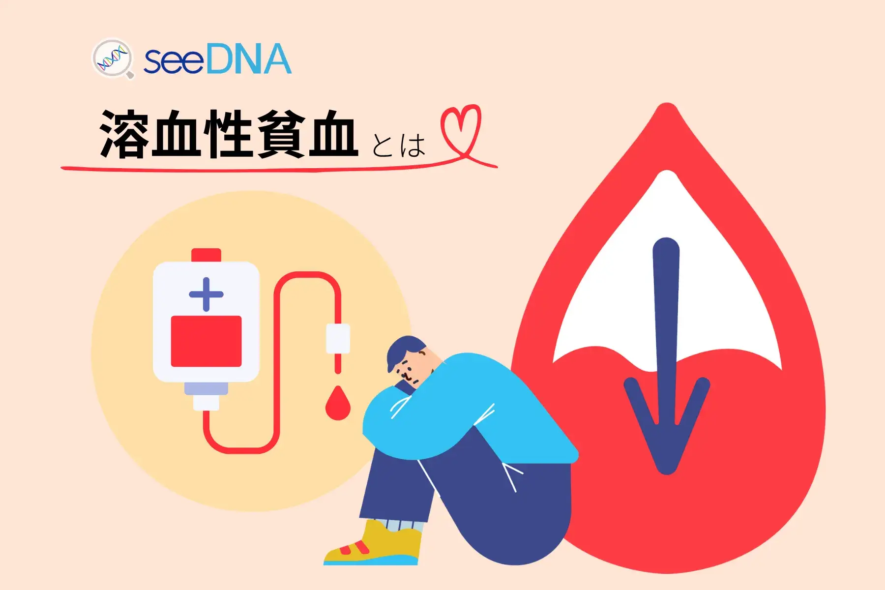
- Hemolytic anemia is a blood disease in which red blood cells are destroyed abnormally quickly.A rapid increase in the number of reticulocytes is a diagnostic indicator.
- Type A mutation in DNA region rs118035855A University of Cambridge study found that people with
- The percentage of reticulocytes in healthy adults is0.5%〜2.5%However, it increases rapidly in hemolytic anemia.
Overview Reticulocyte count or reticulocyte measurement is a test that assesses the number of immature red blood cells in the blood. This is primarily important for diagnosing blood diseases such as hemolytic anemia. Typically, in healthy adults, reticulocytes account for 0.5% to 2.5% of total red blood cells. However, this rate may increase rapidly in hemolytic anemia. Hemolytic anemia is characterized by rapid destruction of red blood cells. At this time, the body tries to manufacture new red blood cells, but at a faster rate, which increases the number of reticulocytes. Therefore, a high reticulocyte count is a suggestive indicator of hemolytic anemia. In patients presenting with symptoms of anemia, high reticulocyte counts may indicate an ongoing hemolytic process. However, additional tests may be needed to determine the root cause of hemolysis. Although the reticulocyte count is an important source of information about the body's response to anemia, other tests are needed for a complete diagnosis. A study by Vuckovic et al. from the University of Cambridge revealed that the risk of developing hemolytic anemia is associated with a DNA region called rs118035855. There are three genotypes in this DNA region: GG, GA, and AA, and it was found that people with the A genotype tend to have a higher risk of hemolytic anemia.
What is hemolytic anemia?
Hemolytic anemia is an anemia that occurs when red blood cells are destroyed (hemolysis) faster than their normal lifespan (about 120 days).When the rate of red blood cell destruction exceeds the rate of new production in the bone marrow, the number of red blood cells in the blood decreases, reducing its oxygen-carrying capacity.
Causes and mechanisms of hemolytic anemia
The cause of hemolytic anemia ishereditary (intrinsic)andAcquired (extrinsic)It is roughly divided into two types.
- Hereditary hemolytic anemia:Red blood cell membrane abnormality (spherocytosis), hemoglobin abnormality (sickle cell disease/thalassemia), enzyme abnormality (G6PD deficiency)
- Acquired hemolytic anemia:Autoimmune hemolytic anemia (AIHA), drug-induced, mechanical hemolysis (prosthetic valve related), infection related
What is reticulocyte testing?
Reticulocytes are immature red blood cells that have just been released into the blood from the bone marrow.Measuring the reticulocyte count is an essential test for diagnosing hemolytic anemia.
- Healthy adults: total red blood cell0.5%〜2.5%is reticulosite
- Hemolytic anemia: The number of reticulocytes decreases because the bone marrow compensates for the rapid destruction of red blood cells by increasing their production.Significant rise
Main symptoms of hemolytic anemia
Symptoms vary depending on the degree and rate of hemolysis.
- chronicFeeling of fatigue/malaise
- jaundice(Yellowing of the skin and eyes)
- Splenomegaly(spleen enlargement)
- Dark urine (hemoglobinuria)
- Palpitations/shortness of breath
Difference between hereditary and acquired
| Comparison items | hereditary hemolytic anemia | Acquired hemolytic anemia |
|---|---|---|
| cause | Genetic abnormalities in the red blood cells themselves | Destruction of red blood cells due to external factors |
| Time of onset | childhood to youth | all ages |
| Representative diseases | Spherocytosis/Sickle Cell Disease | autoimmune hemolytic anemia |
| family history | Yes | Usually none |
| treatment | Spleen removal/blood transfusion | Immunosuppressive therapy/removal of cause |
Diagnostic method
It is diagnosed by the following blood tests.
- Reticulocyte count measurement
- Confirmation of increased indirect bilirubin and LDH levels
- Confirmation of decrease in haptoglobin level
- Direct Coombs test (differentiation of autoimmunity)
- Peripheral blood smear examination
Relationship between genes and hemolytic anemia
Relationship between DNA region rs118035855 and onset risk
A study by Vuckovic et al. (2020) from the University of Cambridge found that the DNA region rs118035855 is associated with the risk of developing hemolytic anemia.
- There are three genotypes of rs118035855: GG, GA, and AA.
- Genotype with type A mutationpeople tend to be at higher risk of hemolytic anemia
Genotype distribution in Japanese (rs118035855)
| Genotype | Percentage of Japanese people | percentage of the world |
|---|---|---|
| GG type | 99.9% | 98.0% |
| GA type | 0.1%以下 | 1.9% |
| AA type | 0.1%以下 | 0.1%以下 |
Rationale for testing
Superficial DNA region: hemolytic anemia
The gene region that most strongly influences hemolytic anemia is rs118035855. The distribution of isomorphic genotypes in Japan is as follows.
- GG
99.9 % - GA
0.1%以下 - AA
0.1%以下
Another gene region involved in hemolytic anemia is rs73225807. The distribution of isomorphic genotypes in Japan is as follows
- TT
99.9 % - TA
0.1%以下 - AA
0.1%以下
Another gene region involved in hemolytic anemia is rs9533095. The distribution of isomorphic genotypes in Japan is as follows
- GG
86.0 % - GT
13.3 % - TT
0.5 %
Basis for inspection
A study by Vuckovic et al. at the University of Cambridge revealed that the risk of developing hemolytic anemia is linked to genes. There is a region called rs118035855 in the human genome, and there are two types of mutations, G and A, in the gene in that region. It was found that people with the type A mutation tend to have a higher risk of hemolytic anemia.
The DNA region investigated this time
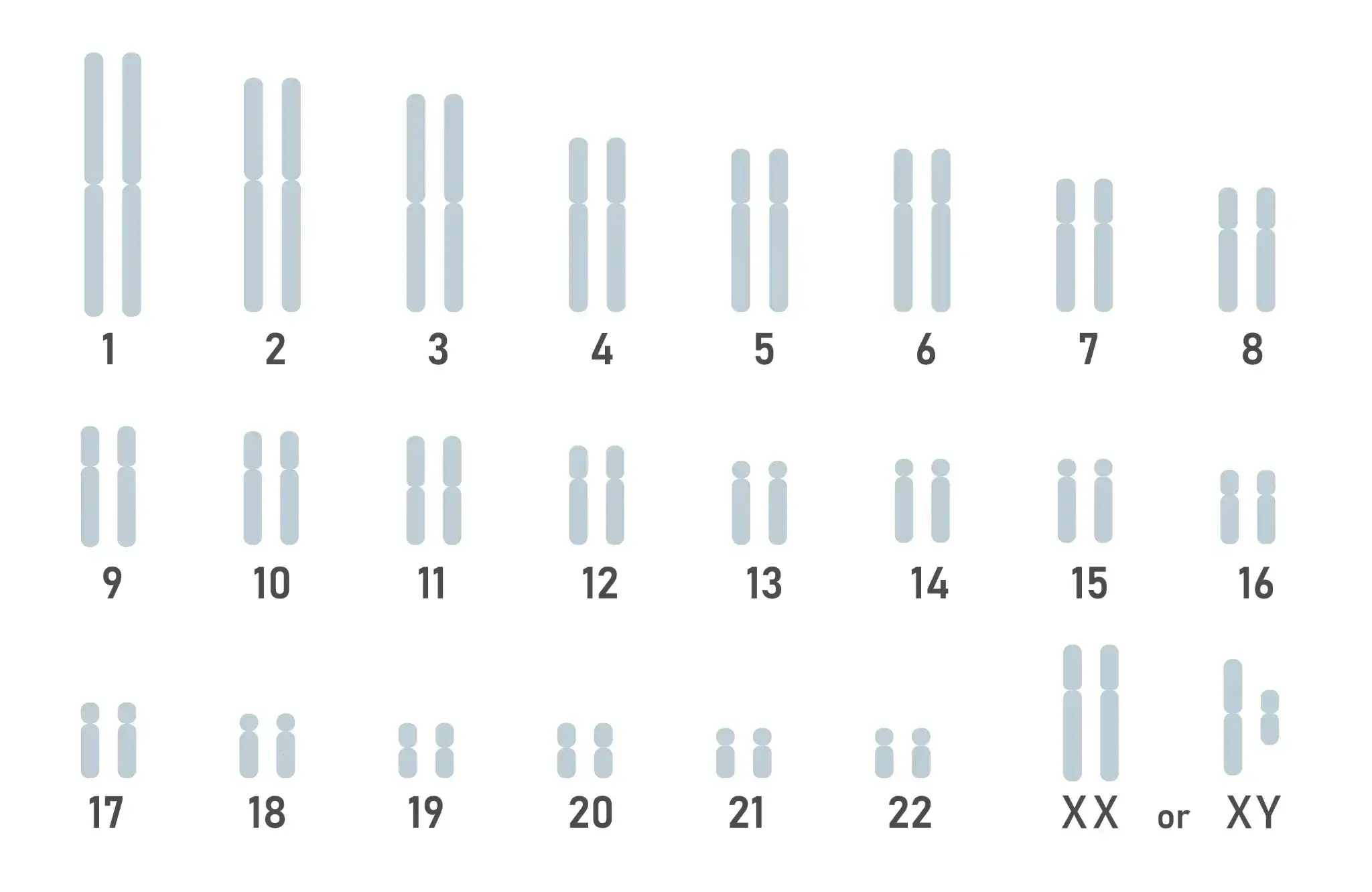
Schematic diagram of DNA map present in cells
- ■
- ■
- ■
- ■
- ■
- ■
- ■
- ■
- ■
Related genes
| Related genes | GREB1L |
|---|---|
| Related genes | RNU6-394P |
| Related genes | LINC02341 |
Frequently asked questions (FAQ)
Q1. What is hemolytic anemia?
Hemolytic anemia is an anemia that occurs when red blood cells are destroyed (hemolysis) faster than their normal lifespan (about 120 days).When the rate of destruction of red blood cells exceeds the rate of production in the bone marrow, oxygen carrying capacity decreases, leading to symptoms such as fatigue, jaundice, and splenomegaly.
Q2. What is the relationship between reticulocyte count and hemolytic anemia?
Reticulocytes (reticulocytes) are immature red blood cellsIt accounts for 0.5% to 2.5% of total red blood cells in healthy adults. In hemolytic anemia, the bone marrow compensates by increasing red blood cell production, so the number of reticulocytes decreases.Significant riseI will. A high reticulocyte count is an indicator of an ongoing hemolytic process.
Q3. What is the cause of hemolytic anemia?
The cause isHereditary (erythrocyte membrane abnormality, hemoglobin abnormality, enzyme abnormality)andAcquired (autoimmune/drug-induced/mechanical hemolysis)It is divided into Carriers of type A mutation in the DNA region rs118035855 tend to be at higher risk.
Q4. Can genetic testing determine the risk of hemolytic anemia?
By examining the genotype of the DNA region rs118035855,Understanding trends in the risk of developing hemolytic anemiaYou can. A study by Vuckovic et al. at the University of Cambridge found that people with the type A mutation tend to be at higher risk.
References
- Reference link 1: 2020 Sep., Dragana Vuckovic, Cell